


OCTavius, the all-in-one ophthalmic solution with 5 essential functions
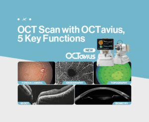
OCTavius integrates five essential diagnostic functions for ophthalmology—3D OCT, Fundus Camera, Angiography, Topography, and Biometry—into a single device.
With its 5-in-1 system, multiple tests can be performed consecutively in one place, allowing patients to undergo examinations comfortably without moving between rooms, while significantly improving clinical efficiency for medical professionals.
In addition, OCTavius minimizes the spatial limitations caused by operating multiple devices separately, maximizing the use of examination room space and creating a more efficient and comfortable clinical environment.
Curious about the five key functions of the OCTavius OCT scan device? In this post, we’ll take a closer look at each feature built into OCTavius and explore the unique benefits they provide!
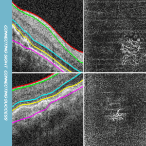
3D OCT : Capture precise 3D images of the macula and choroid in just one second
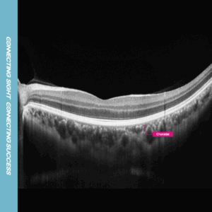
The 3D OCT function of the OCTavius OCT scan device provides rapid and stable scanning, delivering clear images of even the smallest details in the macula and choroid. This makes it highly effective in diagnosing a wide range of ophthalmic conditions, including retinal detachment and vitreous opacity. In addition, with the Enhanced Choroidal Imaging (ECI) mode, signal depth can be inverted to visualize the choroidal layer more distinctly.
Fundus Camera : Produce true-color fundus images with just a single capture
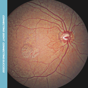
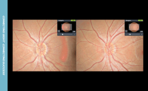
With Huvitz’s proprietary image processing technology, you can obtain true-color fundus images in just a single shot. The system balances brightness evenly—from the dark retina to the bright optic disc —so that arteries, veins, and even fine capillaries are rendered with clarity. Because the colors are free from distortion, fundus images can be analyzed with higher accuracy, ultimately enhancing diagnostic reliability.
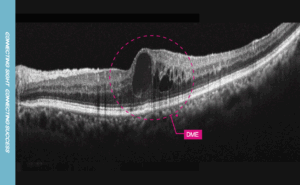
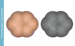
Angiography : Comprehensive scanning of the entire region with fast OCT-A imaging
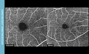
The Angiography function of the OCTavius OCT scan device uses a wide-field mosaic mode, capturing both central and peripheral regions in a single scan. This makes it highly effective for detecting even subtle lesions across a broad area. It also separates the RPEDC (RPE–Drusen Complex) layer with precision, reducing spark noise caused by RPE reflection. As a result, the contours of choroidal neovascularization (CNV) can be visualized with greater clarity.
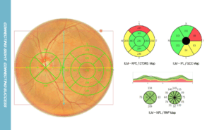
Topography : High-precision analysis of the anterior and posterior corneal surfaces with 16 maps
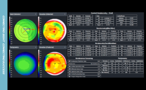
The Topography function analyzes both the anterior and posterior cornea using 16 different maps, including Elevation and Pachymetry maps, to support accurate diagnosis and surgical planning. On a single screen, you can simultaneously view four maps—such as the Axial Map, anterior and posterior Elevation Maps, and Pachymetry Map—making it easy to assess essential parameters like SimK, Meridian, keratoconus indices, and epithelial thickness at a glance.
Biometry : From Precise Measurement to IOL Recommendation
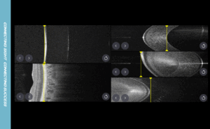
The Biometry function of the OCTavius OCT scan device provides consistent and reliable data through Burst Mode, performing three axial length (AL) scans and five rapid consecutive measurements of central corneal thickness (CCT), anterior chamber depth (ACD), and lens thickness (LT). By combining AL, CCT, CD, and LT values with corneal topography–based K values, the system automatically recommends the most suitable intraocular lens (IOL) for each patient.
OCTavius is an ophthalmic diagnostic device that combines speed, accuracy, and space efficiency in one system. With a single unit capable of performing five essential tests, it enhances not only clinical workflow but also patient satisfaction. More than just an examination device, OCTavius is an all-in-one solution that streamlines the core processes of eye diagnostics—bringing meaningful change to every ophthalmology practice.
