- COMPANY
Looking for more ophthalmology content? Join Huvitz Members now.
Curious about Huvitz?
- BUSINESS
Looking for more ophthalmology content? Join Huvitz Members now.
Curious about Huvitz?
- PRODUCT
Looking for more ophthalmology content? Join Huvitz Members now.
Curious about Huvitz?
- SCIENCE
Looking for more ophthalmology content? Join Huvitz Members now.
Curious about Huvitz?
- PR
Looking for more ophthalmology content? Join Huvitz Members now.
Curious about Huvitz?


MENU
Explore Details
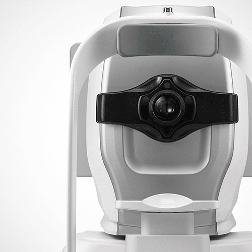
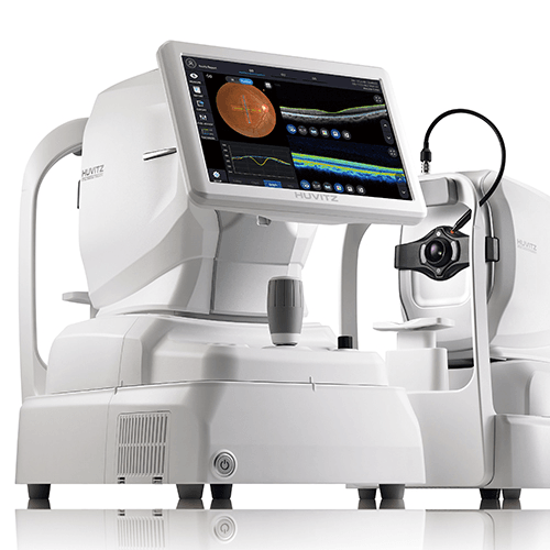
One for All System : 3D OCT, Fundus Camera, Angiography, Biometry, Topography
This is a 5 in 1 optical coherence tomography (HOCT) system capable of performing 3D OCT, Fundus Camera, Angiography, Biometry, and Topography diagnostics and examinations all at once. As an all-in-one system, it enhances efficiency for diagnosing and examining conditions from the anterior to posterior segments of the eye, minimizing consultation time and maximizing space utilization.
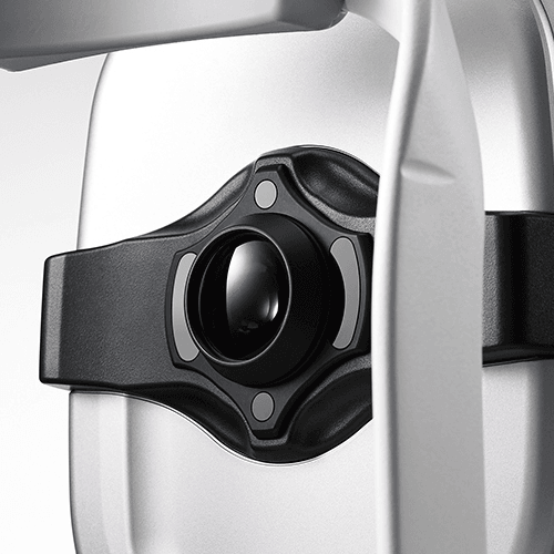
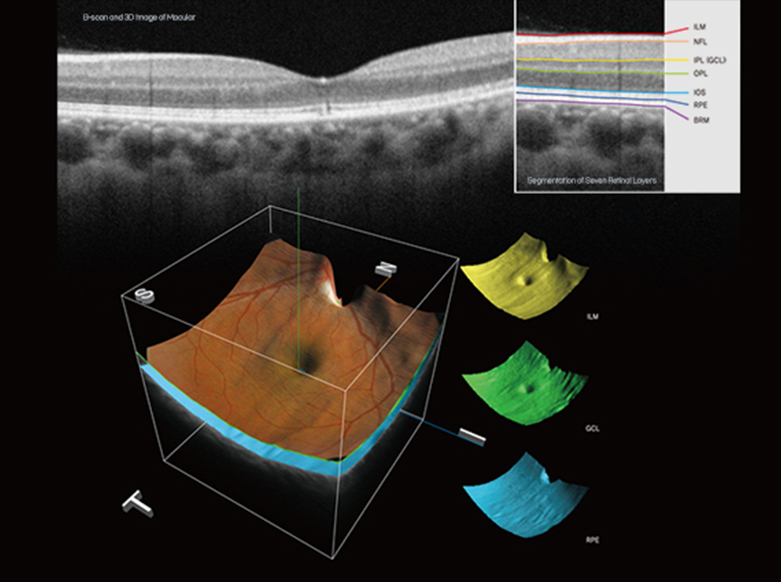
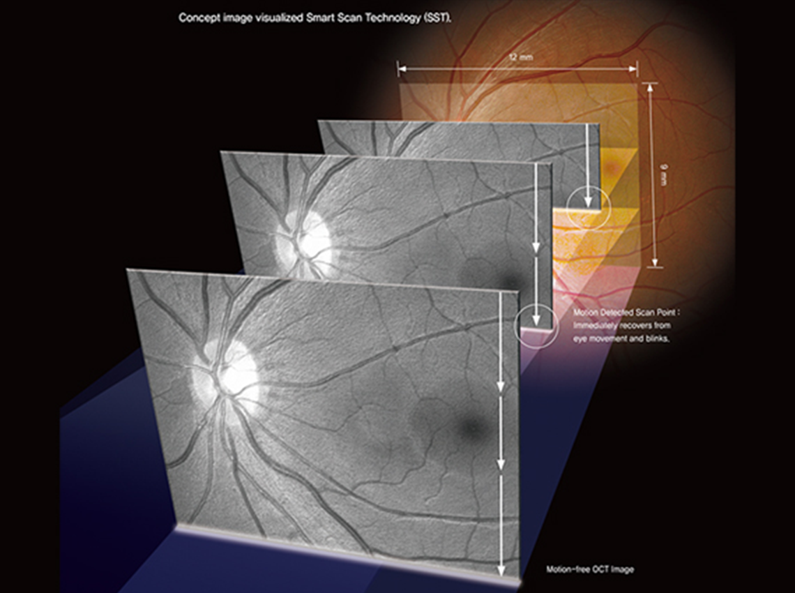
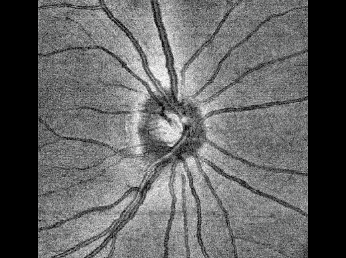
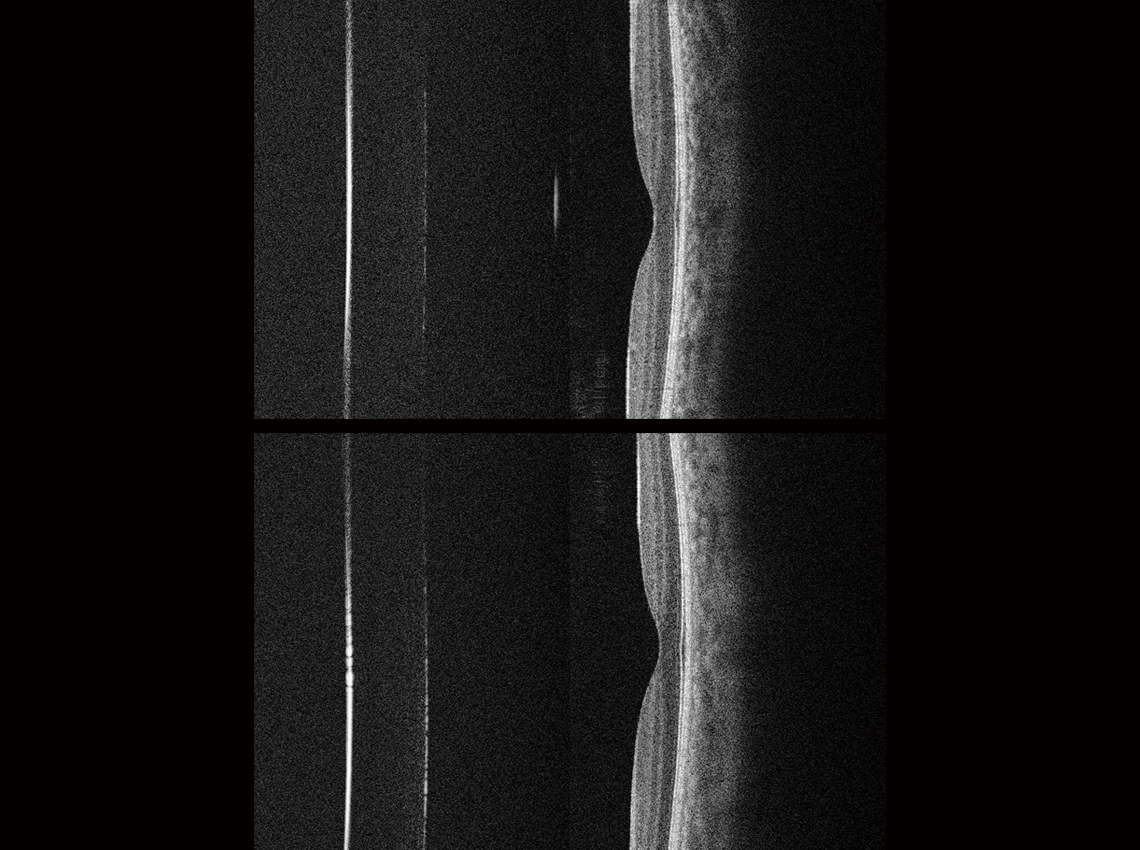
High-Speed & High-Quality
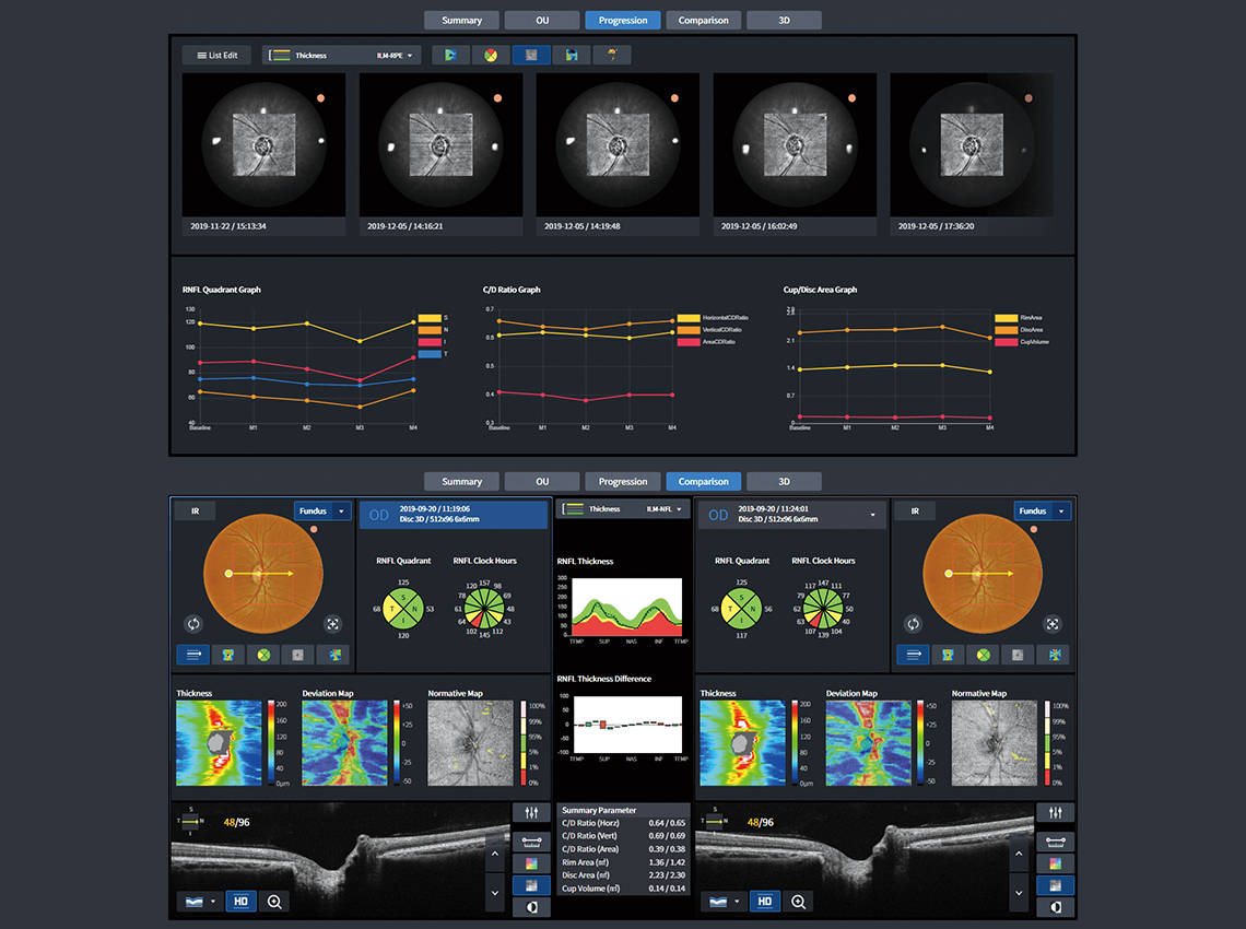
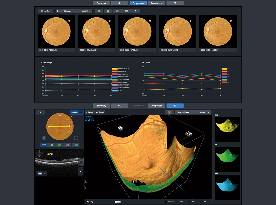
Huvitz's optical technology and image software deliver rapid scanning and high-quality imaging. With an astonishing speed of 80,000 A-scans per second, coupled with high-resolution images, it vividly displays a wealth of information such as the three-dimensional structure of the retina, thickness, and differentiation of the macula.
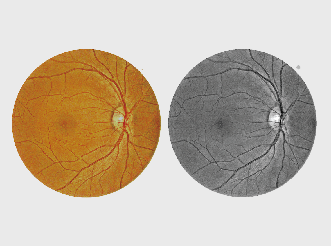
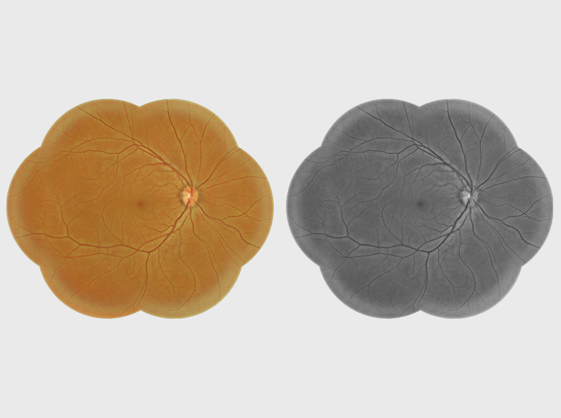
Full Color Fundus Image
Acquiring high-resolution, contrast-adjusted color retinal images is invaluable for analysis and clinical diagnosis of the posterior segment of the eye. We have combined low flash intensity, rapid capture speed, quiet operation, small pupil mode, and automatic blink detection to capture the finest images possible.
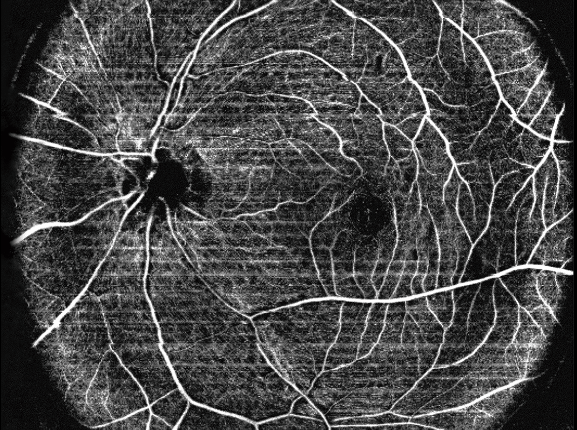
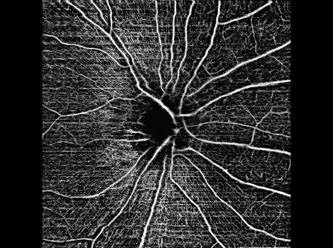
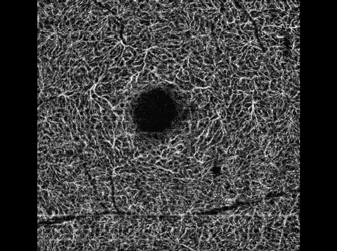
Innovative Angiography
Huvitz's own optical technologies, Real Time Tracking / Noise Cancelling / Motion Correction work together to automatically analyze and visualize the Retina, Microvasculature of Choroid.
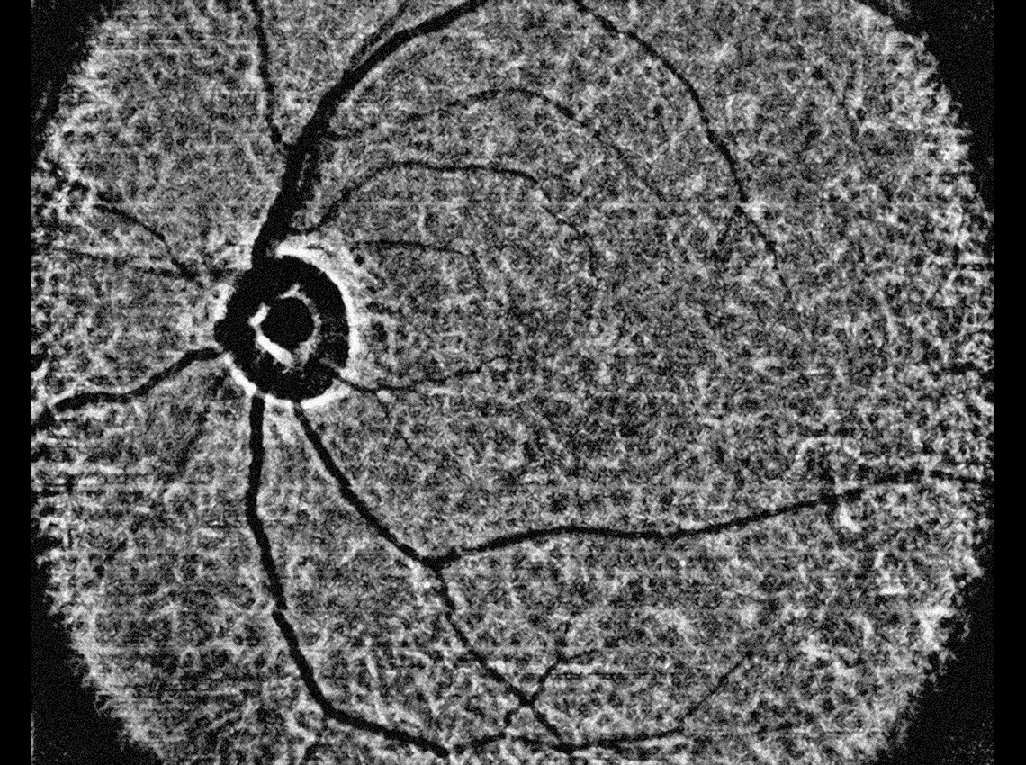
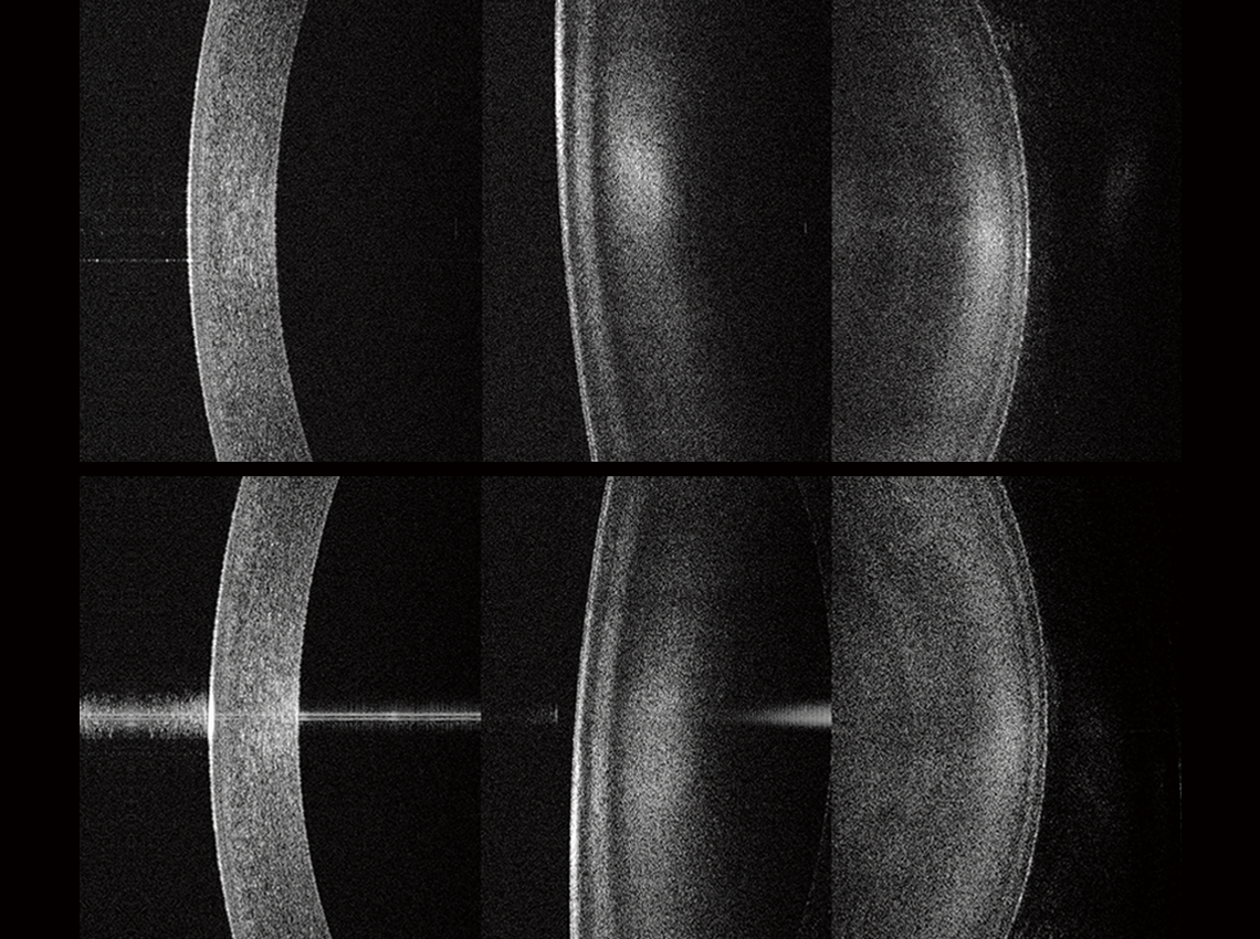
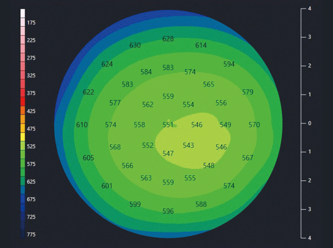
Detailed measurement of biometry and topography
From the cornea to the macula, HOCT displays 2D images and provides all data along the anterior and posterior segments. After measurement is complete, the user can identify and make adjustments where necessary.
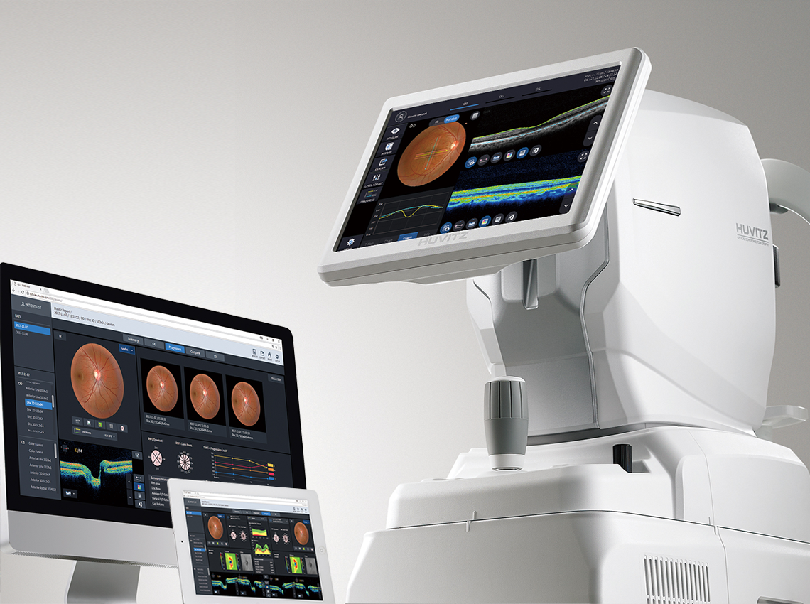
Accurate Analysis
Through integrated analysis, it's possible to grasp personalized symptoms, diseases, and their progression for each patient at a glance. This includes tracking pathological changes with Progression analysis, comparing pre- and post-treatment states with Compare functionality, analyzing pathological conditions from various perspectives such as key indicators compared to normative data
Video
Reviews