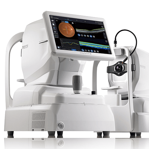
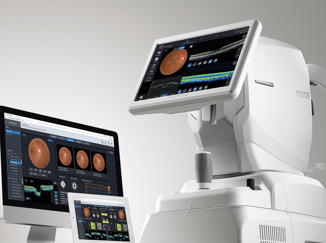
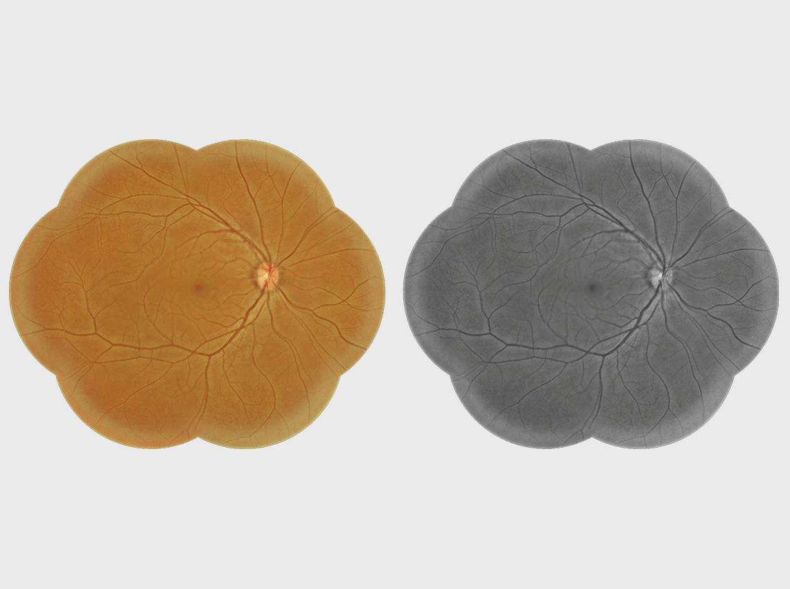
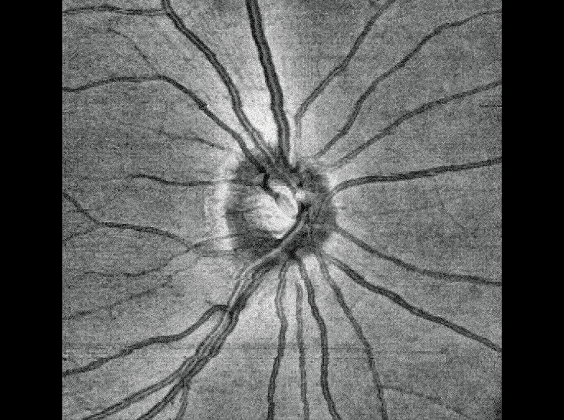
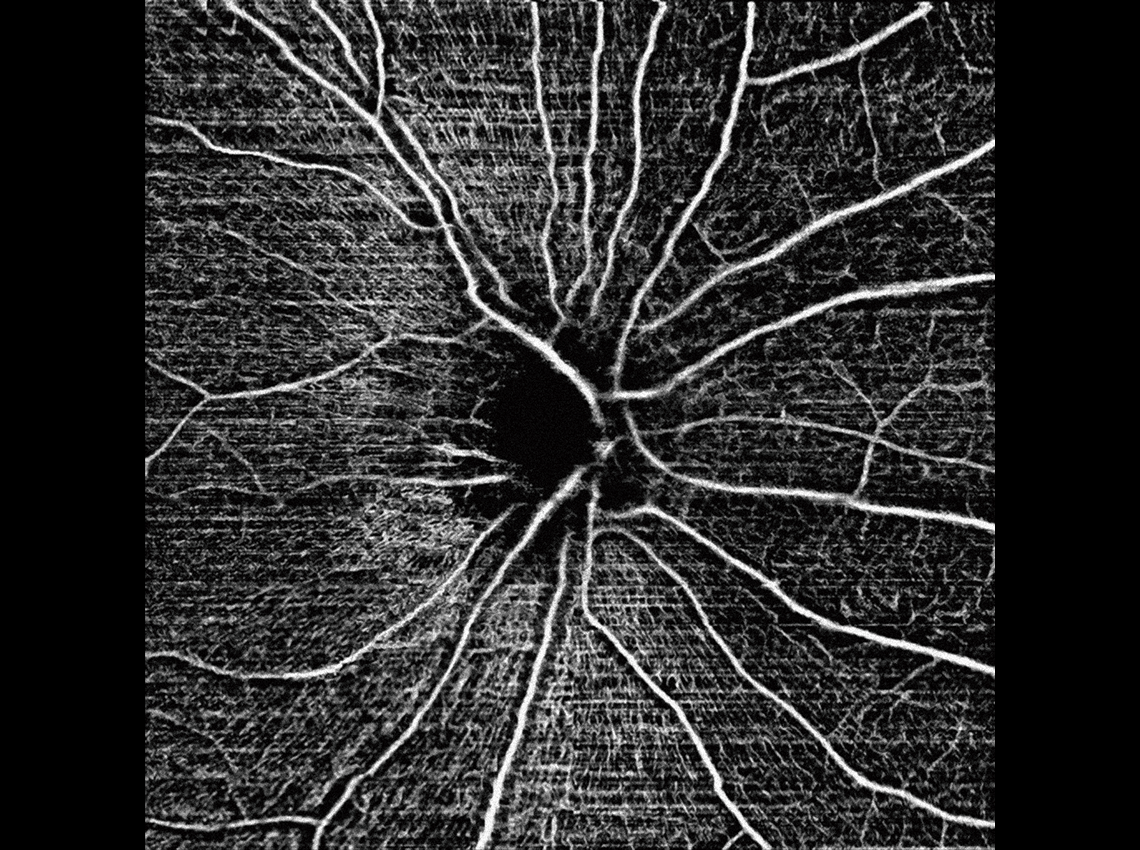
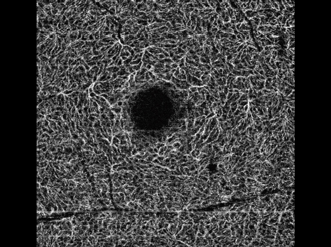
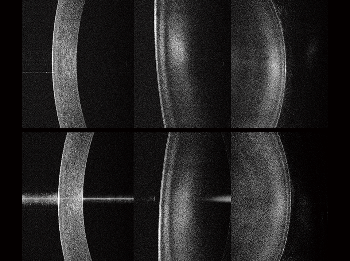
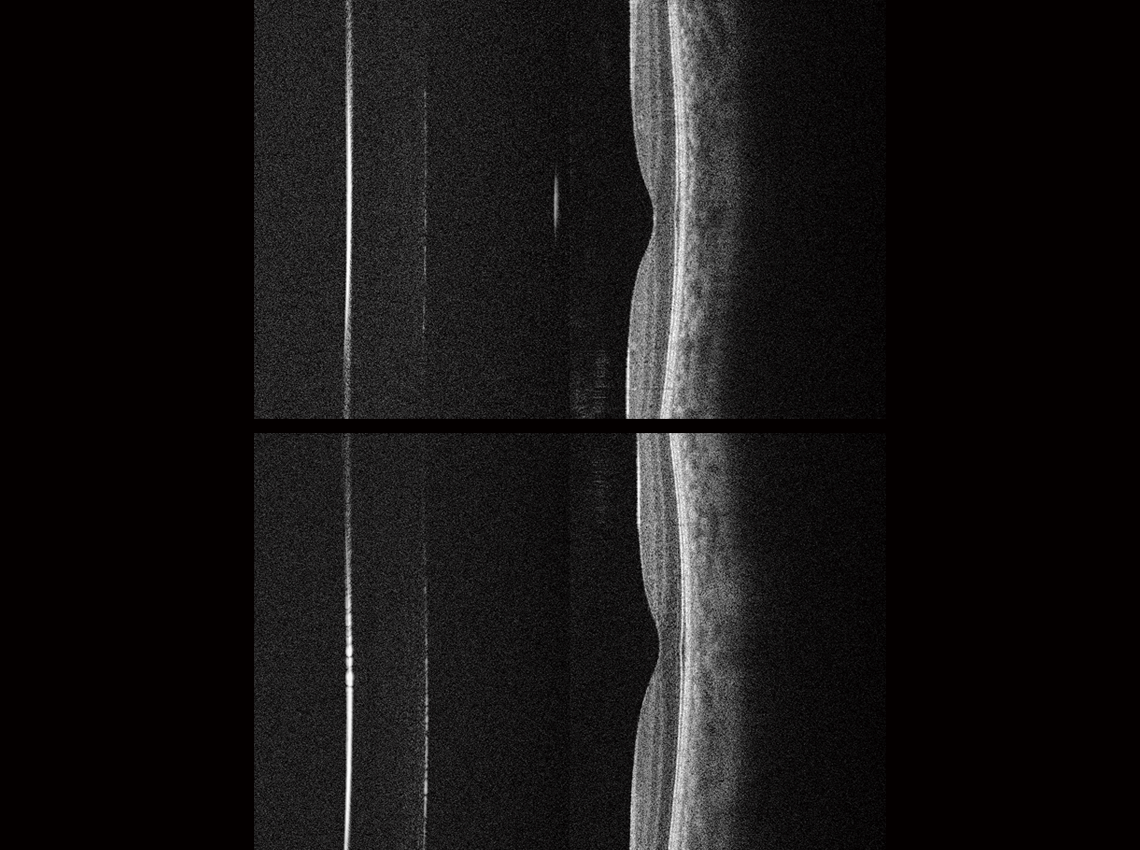
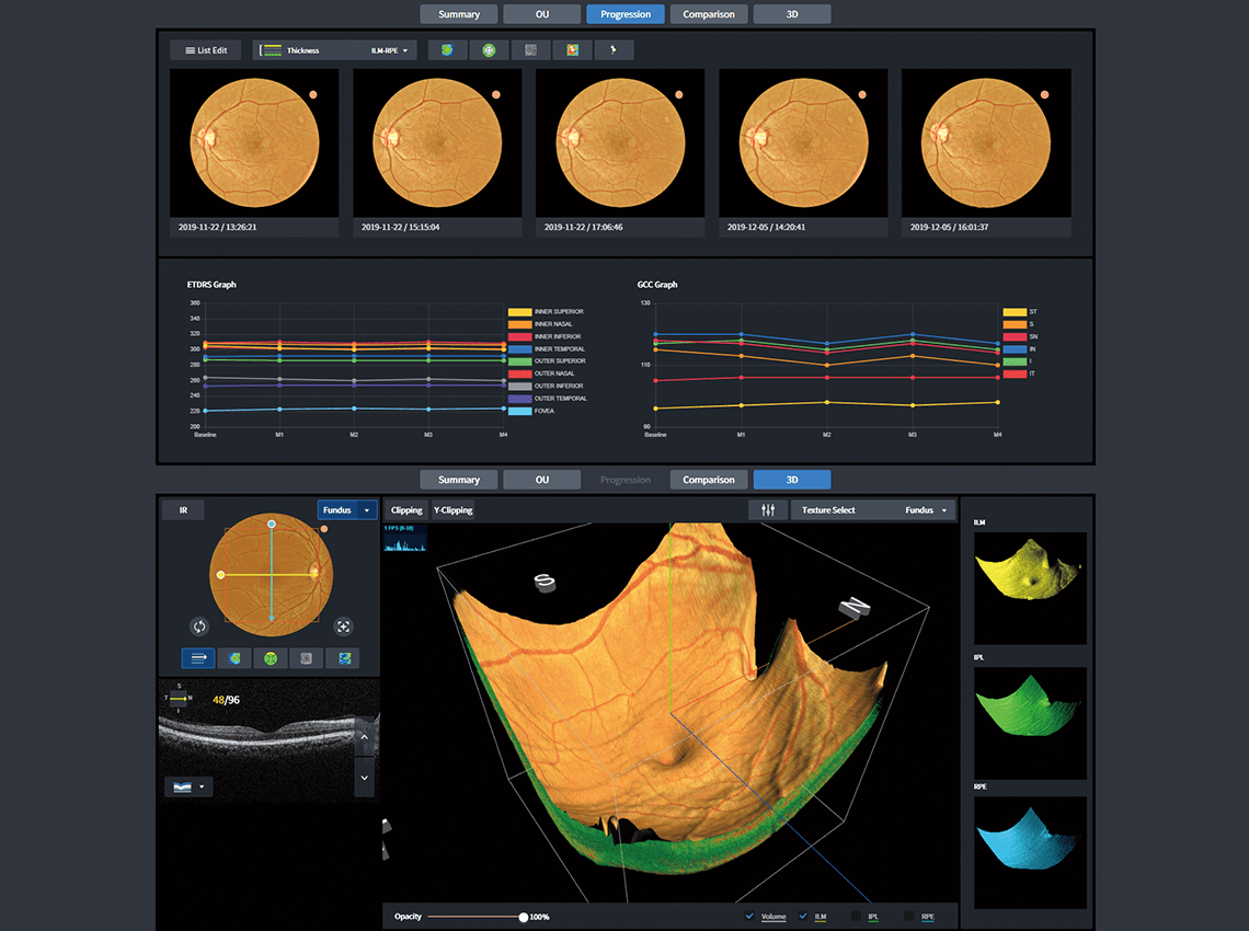
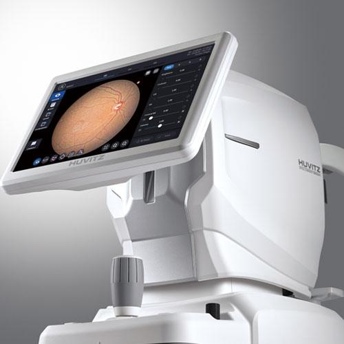
- COMPANY
Looking for more ophthalmology content? Join Huvitz Members now.
Curious about Huvitz?
- BUSINESS
Looking for more ophthalmology content? Join Huvitz Members now.
Curious about Huvitz?
- PRODUCT
Looking for more ophthalmology content? Join Huvitz Members now.
Curious about Huvitz?
- SCIENCE
Looking for more ophthalmology content? Join Huvitz Members now.
Curious about Huvitz?
- PR
Looking for more ophthalmology content? Join Huvitz Members now.
Curious about Huvitz?
MENU
Explore Details
One for All System : 3D OCT, Fundus Camera, Angiography, Biometry, Topography
HOCT 하나로 3D OCT, Fundus Camera, Angiography, Biometry, Topography 검사 및 진단이 한번에 가능한 5 in 1 망막단층진단기입니다. 올인원 시스템으로 전안부부터 후안부 질환 진단 및 검사를 위해 효율성을 높이고, 진료시간 단축 및 공간활용을 극대화하였습니다
빠른 스캐닝과 고화질의 이미지 제공
휴비츠의 광학기술과 이미지 소프트웨어는 빠른 스캐닝과 고품질 이미지를 제공합니다. 80,000 A-scan/sec.의 속도와 고화질 이미지로 Retina의 3차원 구조, Macula의 두께 및 구분 등 많은 정보를 생생하게 보여줍니다.
Full Color Fundus Image 획득
High-resolution, Contrast가 조율된 Color Retinal Images를 획득함으로써 Posterior Segment of Eye에 대한 분석과 임상진단에 유용합니다. 낮은 플래시 강도, 빠른 캡처 속도, 조용한 작동, Small Pupil Mode 및 자동 깜박임 감지 등 최상의 이미지를 캡처하기 위해 모두 모았습니다
Angiography 기술로 망막 및 맥락 미세혈관 분석 및 시각화
안구의 미세한 움직임을 추척하여 측정오류를 줄여주는 Real Time Tracking, 명암의 디테일을 섬세하게 다듬어 시각화하는 Noise Cancelling, 혈관의 틀어짐을 보정하는 Motion Correction이 상호 연동하여 망막 및 맥락막의 미세혈관을 자동분석하고 시각화 합니다.
Biometry와 Topography의 세부적인 정보 측정
각막 정점부터 황반까지 그래프가 아닌 2D 이미지로 보여주고, 눈의Anterior&Posterior Segment의 모든 데이터를 제공합니다. 또한 각막 전/후면의 곡률과 두께를 측정하여 각막의 전면과 후면을 포함한 16개의 맵을 제공하여 보다 정확한 분석을 할 수 있습니다.
다양하고 세분화된 정보 제공으로 정확한 분석 결과 제공
통합된 분석으로 환자마다 특성화된 증상, 질병 그리고 그 진행과정을 한눈에 파악할 수 있습니다. 병리적 변화를 추적해 보는 Progression, 전과 후의 상태를 비교하는 Compare, Normative Data와 비교되는 주요 지표값 등 다양한 관점에서 병리 상태를 분석합니다. 또한 빠른 개요부터 단순 비교, 복합 평가까지 의미 있는 보고서를 완료합니다.
