
HIS-5000
Huvitz Imaging System
Imaging system for capturing images and videos observed through a slit lamp
- Utilizing a 1-inch image sensor for superior image and video capture
- Analyzing measurement data
- Simplifying patient management
MENU
Explore Details
Utilizing a 1-inch image sensor for superior image and video capture
The use of a 1-inch image sensor enables capturing superior quality images and videos. With the Live Tool graphic library, images in formats like JPG, TIFF, and RAW can be adjusted for brightness, contrast, color channels, saturation, inversion, sharpness, and red-free effects
Analyzing measurement data
Various graphic effects such as zooming, scaling, and rotation can be applied to selected examination images for comparative observations. Additionally, an animation feature allows for observing the progression and transition of diseases across multiple examination images from different time points.
Simplifying patient management
Measured data in standard formats like DICOM and HIIS-1 can be viewed on a PC. The HIS system, based on MS Access Database, allows easy management of examination information, including diagnostic records, examination histories, and other content. Capturing and editing images and videos facilitate the generation and sharing of patient data. From data generation to progress monitoring, patient management becomes more streamlined
Video
Huvitz MEMBERS
더 많은 안과컨텐츠를 원하신다면
휴비츠멤버스에 가입하세요.
Specifications
1. Digital camera
Camera Model
HIS-5000U(10.0M)
HIS-5000U(5.0M)
Image Sensor
1/2" CMOS
1" CMOS
Image Size
up to 3840 x 2748 pixels
up to 2592 x 2048 pixels
Cell Size
1.67μm x 1.67μm
4.65μm x 4.65μm
Transmit Method
USB 2.0
USB 3.0
Transmit Speed
480Mbps
5.0Gbps
Frame Rate
Maximum 30fps
Lens Mount
C-Mount
Shutter
Rolling Shutter
Global Shutter
Photographing
External Trigger or Software Trigger
Power Consumption
5V DC, 2.5W
5V DC, 3.5W
2. System requirements
CPU
Intel® Core™ i3 (2.4GHz or over)
Intel® Core™ i5 (3.4GHz or over)
Memory
Over 2GB recommended
Over 4GB recommended
Video Card
On board graphics chip
(PCI/PCIe graphics card with DirectDraw support DirectX 9.0 or Higher Recommended)
System
Microsoft® Windows®7 SP1, Windows®8, Windows®10
Monitor
Resolution 1920 x 1080 Recommended
* Specification and design are subject to change without notice. * The PC and table are optional.
Related Products
HT-5000
Applanation Tonometer
HS Series
Slit Lamp
HNT-1/1P
Non-contact Tonometer
HFC-1
Non-Mydriatic Fundus Camera
HTR-1A
Auto Ref/Keratometer, Non-contact Tono/Pachymeter
HTG-1
Full Corneal Optical Topographer
HIIS-1
Huvitz Integrated Data Management System
HBM-1
Optical Biometer with Full Corneal Topography System
HOCT-1/1F
Optical Coherence Tomography
FAQ
Q. What is the HIS-5000 imaging system?
A. The HIS-5000 is Huvitz’s dedicated imaging system designed to capture high-quality images and videos observed through a slit lamp. Equipped with a 1-inch image sensor, it delivers clear diagnostic images and supports various forms of data evaluation, including comparative analysis and progression tracking.
Q. What comparison observation features are supported by the HIS-5000?
A. The HIS-5000 enables precise comparative observation by applying various graphic functions—such as zooming, panning, and rotation—to selected examination images. In addition, its animation feature, which links images captured at different time points, allows clinicians to intuitively track disease changes and progression. Furthermore, reference images can be registered and retrieved by disease category, enabling efficient comparison with standard reference images during diagnosis.
Q. What functions are offered by the HIS-5000 imaging system?
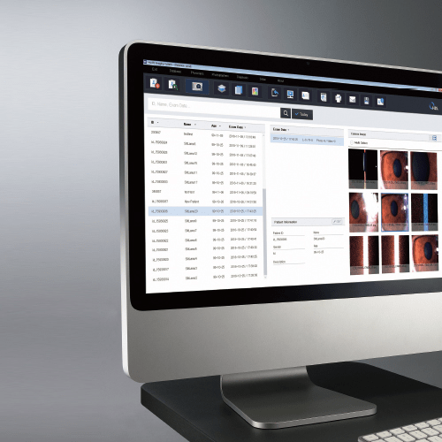
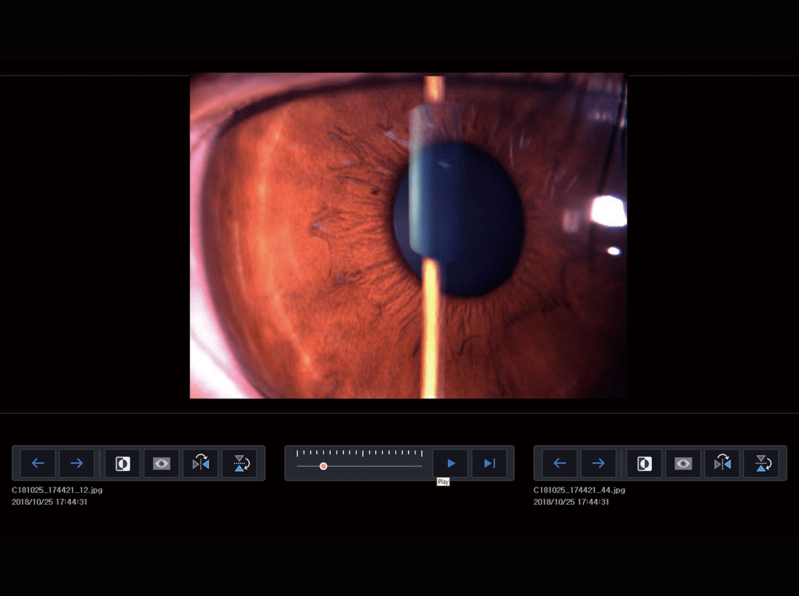
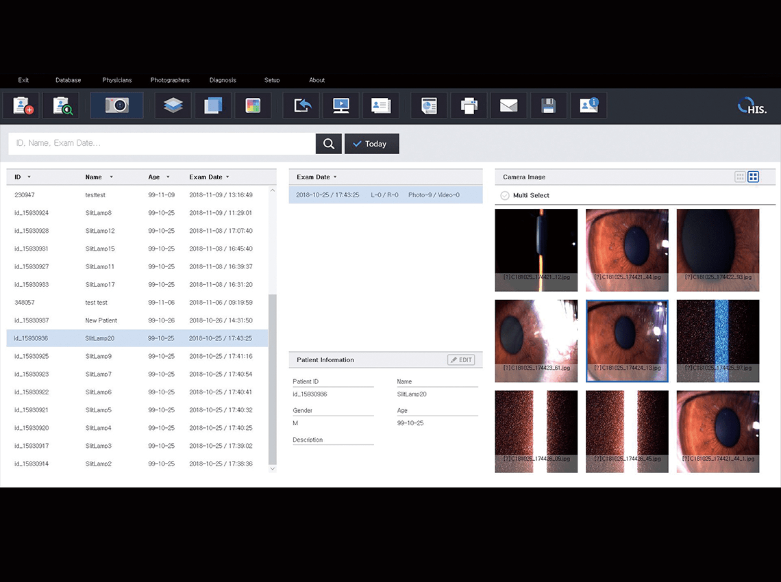
A. The HIS-5000 supports magnification, zooming, rotation, and time-sequence animation of slit lamp images to facilitate comparative observation and progression analysis. Integration with DICOM and HIIS-1 enables seamless management of examination data on a PC. With its image and video capture and editing capabilities, the system enhances efficiency across diagnosis, documentation, and long-term patient monitoring. In addition, the Time Machine and Flicker functions allow users to select the optimal image from multiple frames captured in a single shot, ensuring high-quality images without the need for re-acquisition.
Q. In which clinical environments is the HIS-5000 most suitable?
A. The HIS-5000 is ideal for clinical settings where detailed observation and documentation of anterior segment conditions—such as keratitis, conjunctivitis, corneal epithelial defects, and cataracts—are essential. Its high-resolution image·video capture and time-based comparison tools help clinicians track disease progression and treatment response. The system also enables efficient management of patient information and examination history, supporting patient consultation and follow-up care.
Q. How does the HIS-5000 digitize slit lamp observation data?
A. The HIS-5000 captures high-quality images and videos observed through the slit lamp and stores them as digital data. This enhances the accuracy of clinical documentation and is highly useful for patient communication, follow-up comparisons, and evaluating changes in disease progression.


