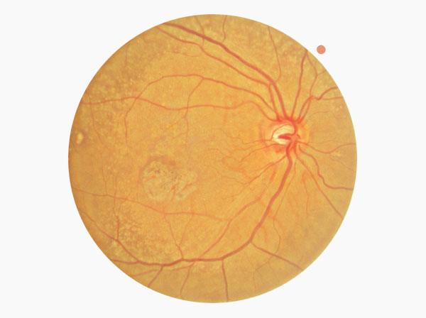
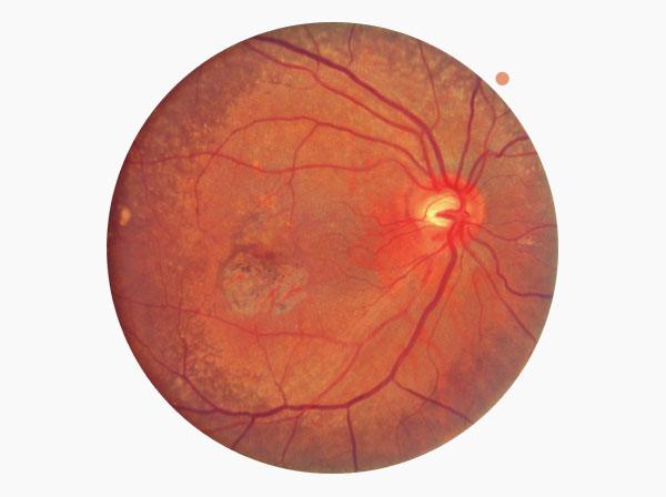
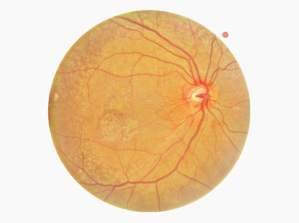
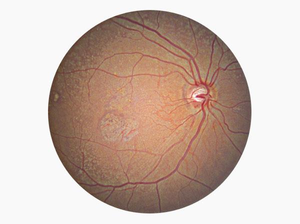
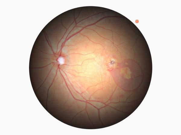
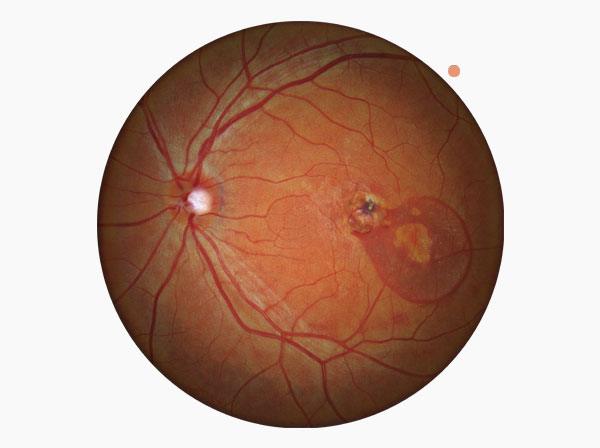
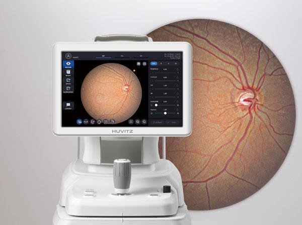
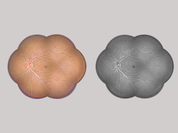
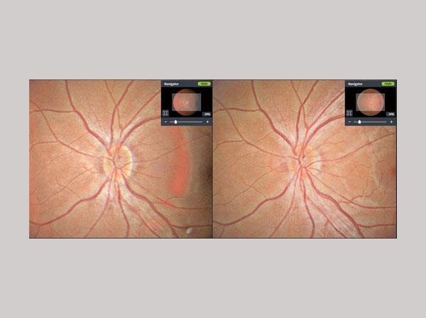
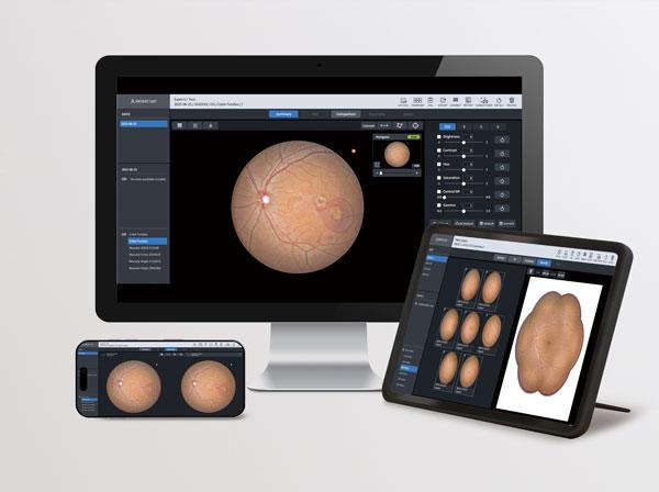
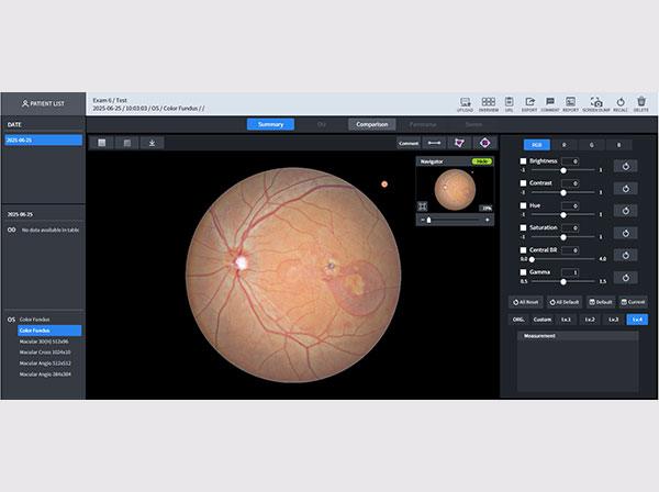
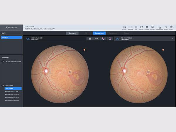
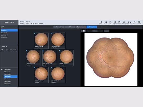

HFC-1
Non-Mydriatic Fundus Camera
PC-integrated fundus camera delivering clear and distortion-free images in a single shot
- User-selected fundus color mode
- Providing clearer fundus images through brightness and color adjustment
- Wide-ranging visualization of lesion location and extent without distortion
- Accurate comparative views based on detailed information
MENU
Explore Details
User-selected fundus color mode
Four types of fundus color images are provided, allowing users to choose the optimal color tone according to their diagnostic style or visual preference for precise observation of blood vessels.


Enhanced lesion visibility through brightness and color adjustment
By adjusting the Central BR and Gamma values, the brightness and color of the fundus image can be finely tuned. This allows users to observe specific areas where lesions are located more clearly.
True Color Fundus in One Shot
With 12-bit color depth and gamma correction, it captures true color fundus images in a single shot—free from color distortion. It accurately balances contrast across both dark retinal areas and bright optic discs, delivering clear visualization of arteries, veins, and even fine microvasculature. Additionally, it automatically aligns and merges multiple fundus images—up to seven—to generate a widefield panoramic view, enabling intuitive identification of lesion location and extent in a single, comprehensive image.


Accurate comparative views based on detailed information
Through detailed and comprehensive analysis, it is possible to grasp personalized symptoms, diseases, and their progression for each patient. This includes comparing pre- and post-treatment states with Comparison, and analyzing pathological conditions from various perspectives such as Single and Panorama reports.
Huvitz MEMBERS
더 많은 안과컨텐츠를 원하신다면
휴비츠멤버스에 가입하세요.
Specifications
1. Technical Specifications
Type
Non-mydriatic fundus camera
Resolution
Center: 60 lines/mm or more
Middle (r/2): 40 lines/mm or more
Peripherally: 25 lines/mm or more
Angle of view
45°
Camera
Color Image Sensor
Minimum pupil diameter
4.0mm (Normal mode), 3.3mm (Small pupil mode)
Flash light
White light
Working distance
33mm
Dioptric compensation for patient’s eye
-33D ~ +33D total
-33D ~ -7D with minus compensation lens
-13D ~ +13D with no compensation lens
+7D ~ +33D with plus compensation lens
Auto tracking
X, Y for positioning, Z for working distance
2. Hardware Specification
Display
12.1 inch, 1280x800 pixel, Touch panel color LCD
Internal fixation target
LCD (internal), White LED (external)
Power supply
AC 100~240V, 50/60Hz, 1.6~0.7A
PC
Built-in computer
LCD Tilting angle
70˚
Dimensions
330(W) x 542(D) x 521(H)mm
Mass
28kg
3. Measurement Range
Horizontal movement
70mm (back and forth), 100mm (left and right)
Vertical movement
30mm
Chinrest movement
62mm (up and down), motorized
* Specification and design are subject to change without notice.