


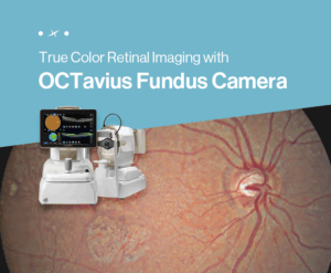
See the Signs of Diabetic Retinopathy in True Color with OCTavius
As the aging population accelerates, the prevalence of chronic ophthalmic diseases such as diabetic retinopathy and age-related macular degeneration (AMD) continues to rise. Consequently, fundus imaging has become an essential examination in ophthalmic practice and is now recognized as a key tool for precise diagnosis and prognosis management.
In modern ophthalmology, it has become increasingly common to combine fundus photography using a Fundus Camera with OCT (Optical Coherence Tomography). By leveraging these two technologies together, clinicians can achieve deeper visualization and a more comprehensive understanding of lesion location and color, enabling a more three-dimensional diagnostic environment.
In response to this growing demand for diagnostic precision, Huvitz has introduced OCTavius, an integrated solution that combines a true-color based Fundus Camera with OCT. This innovative system enhances both diagnostic accuracy and clinical efficiency.
In this article, we will take a closer look at the unique features and advantages of the OCTavius Fundus Camera.
True-Color Fundus Images in One Shot
For accurate diagnosis, it is essential to visualize the location and extent of lesions without distortion and over a wide field of view. The OCTavius Fundus Camera, powered by Huvitz’s proprietary Smart Viewing Technology (SVT), provides high-quality true-color fundus images with precise color accuracy for reliable clinical assessment.
√ Capture the finest retinal microvasculature with uncompromised detail
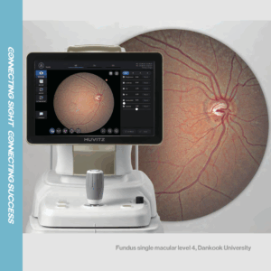
With 12-bit color depth and Gamma correction technology, OCTavius captures true-color fundus images in a single shot. Brightness distribution is balanced to represent both the dark retina and the bright optic nerve head, enabling clear visualization of arteries, veins, and even microvasculature.
√ Wide-angle panorama view for intuitive lesion monitoring
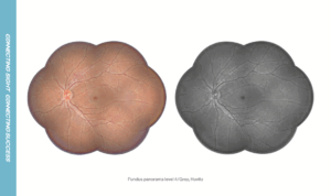
By combining 2 to 7 fundus images, OCTavius generates a wide-angle panorama view of the retina. This allows clinicians to observe the entire fundus structure in a single image, making it particularly effective for diagnosing and monitoring diseases with wide lesion coverage, such as diabetic retinopathy.
√ Structural analysis of the optic nerve head
Images captured from different left and right viewpoints enable precise analysis of the structural characteristics of the optic nerve head, supporting the diagnosis of diseases such as glaucoma.
√ AI-based analysis of 12 retinal lesions
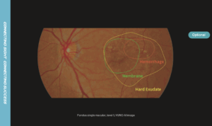
The AI-based fundus analyzer divides the retina into eight regions, detects a total of 12 lesions, and accurately displays their exact locations. This feature improves both diagnostic efficiency and accuracy, enhancing the overall clinical workflow.
Advanced Fundus Imaging with User-Selected Color Modes
The way fundus images are displayed can vary depending on the clinician’s diagnostic style and visual preference. OCTavius provides advanced customization features that allow users to adjust brightness, color tone, and visualization settings, delivering images optimized for individual diagnostic needs.
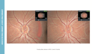
√ User-selected fundus color mode
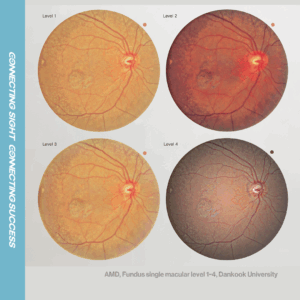
The True Color Fundus feature offers four levels of color modes (Level 1–4), enabling users to select the optimal color tone for their diagnostic objectives and visual preference.
√ Enhanced lesion visibility through brightness and color adjustment
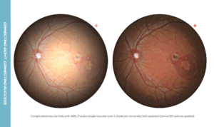
By adjusting the Central BR and Gamma values, users can finely control the brightness and color of the fundus image. This allows for clearer observation of specific lesion areas, improving diagnostic accuracy.
√ RGB color channels for lesion-focused analysis
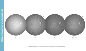
Depending on the lesion characteristics, users can select R (Red), G (Green), or B (Blue) channels to highlight targeted lesion areas and observe them in greater detail.
[Clinical Case] Real-World Diagnostic Examples Using OCTavius
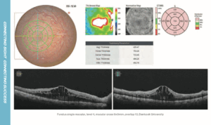
Case 1: Wet Age-Related Macular Degeneration (Wet AMD)
– Findings: In Wet AMD, choroidal vessel dilation is observed, accompanied by hemorrhagic pigment epithelial detachment (PED) and compression of the choroidal capillaries.
– Recommended Treatment: Accurate diagnosis is supported through OCT and fundus photography, and intravitreal injection therapy is recommended.
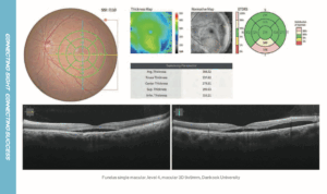
Case 2: Central Retinal Vein Occlusion (CRVO)
– Findings: In CRVO, severe macular edema is observed in the central region, accompanied by inner retinal structure damage, venous dilation, and retinal hemorrhage.
– Recommended Treatment: Conduct visual acuity testing and fluorescein fundus angiography, followed by intravitreal injection or steroid therapy as needed.
OCTavius is an integrated solution that combines a true-color based Fundus Camera with OCT in a single device, enabling:
– 3D visualization of lesion location and extent
– Accurate prognosis monitoring of disease progression
– Reduced diagnostic time, enhancing both patient satisfaction and clinical efficiency
👉 Discover more features of OCTavius.
