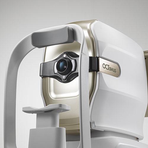
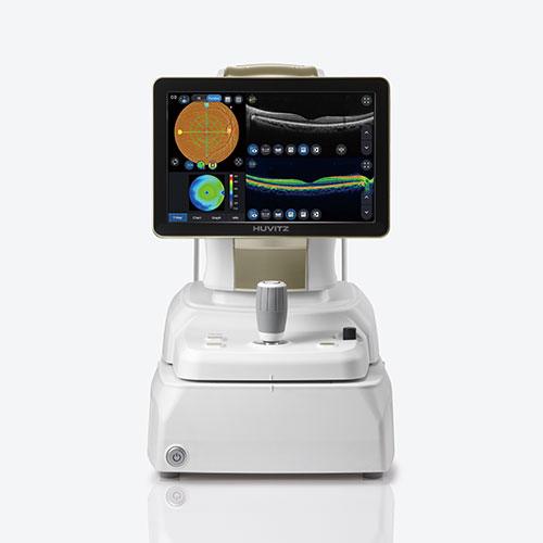
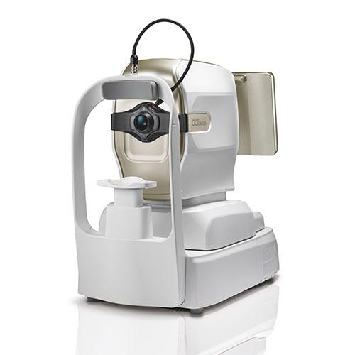
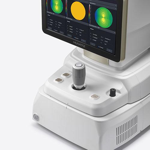
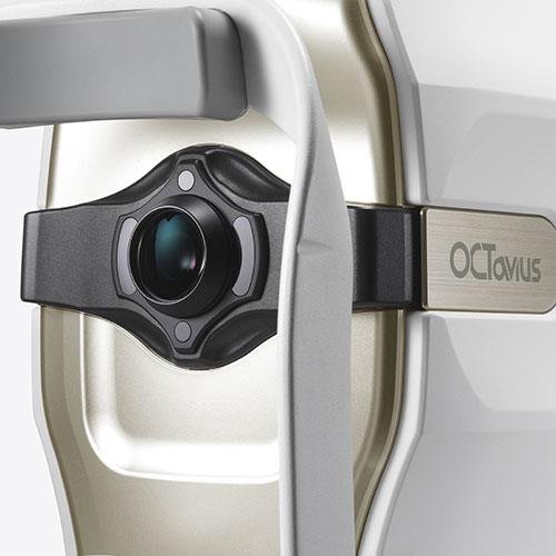
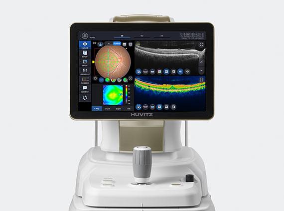
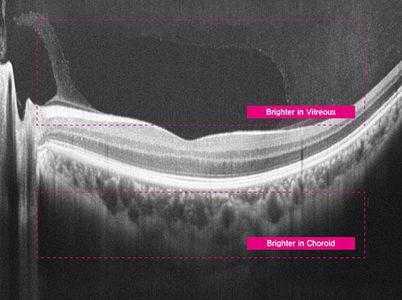
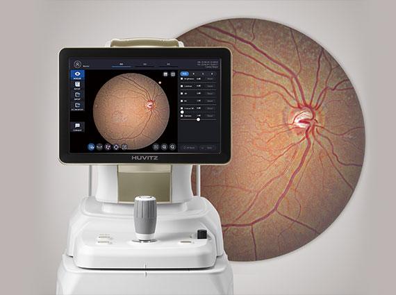
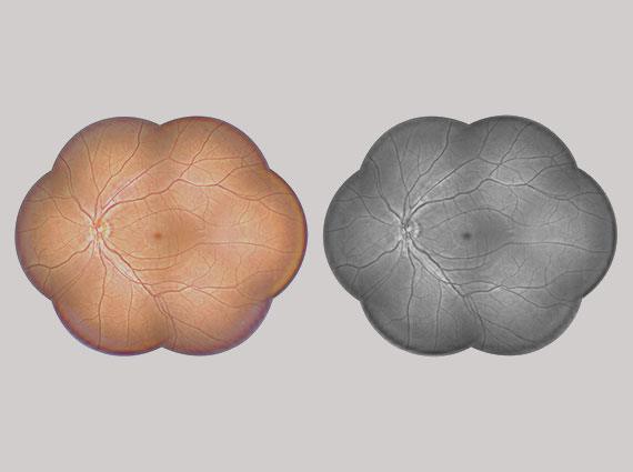
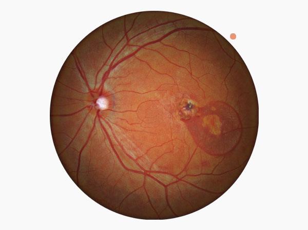
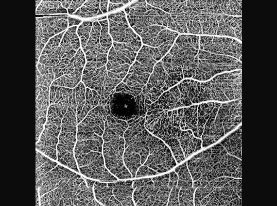
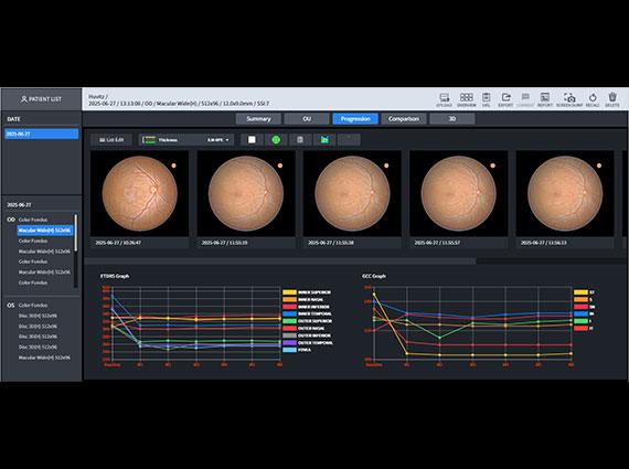
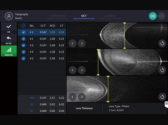
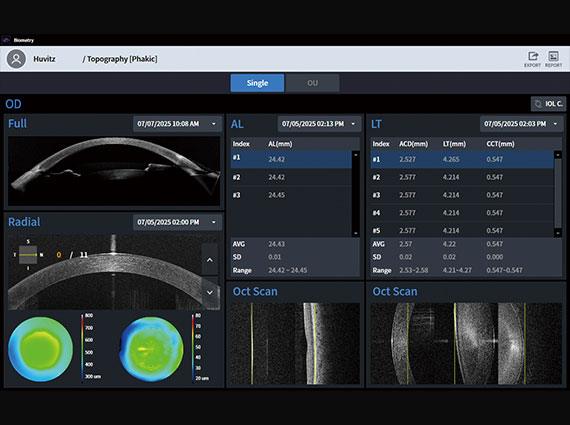
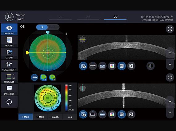
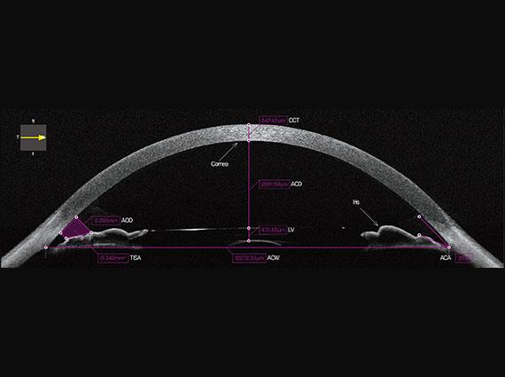
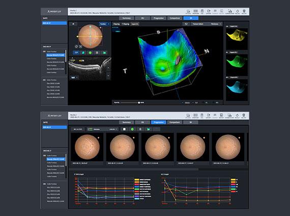
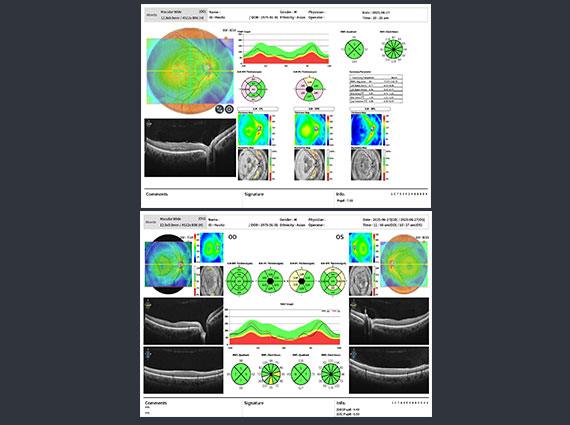
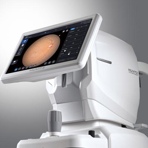
Check out Huvitz's Global Partners

OCTavius

Optical Coherence Tomography

A 5 in 1 ophthalmic diagnostic solution with rapid 80,000 scans per second and true color image quality

Explore Details

5 in 1 System : 3D OCT, Fundus Camera, Angiography, Biometry, Topography

OCTavius integrates five essential diagnostic functions—3D OCT, Fundus Camera, Angiography, Biometry, and Topography—into a single device. It offers seamless operation and delivers precise results needed for clinical decision-making. With the 5-in-1 system, enhance your diagnostic accuracy while maximizing workflow efficiency and exam room space.

80,000 A-scan/sec
Quick Capture, Clear Answer

Even with involuntary eye movements or blinking, OCTavius captures stabilized, high-resolution 3D images through ultra-fast scanning at up to 80,000 A-scans per second. This ensures clear, repeat-free imaging—ideal even for beginners. Visualized retinal layers allow clinicians to observe pathological structures such as layer thickness and macular differentiation with greater clarity and confidence.

True Color Fundus in One Shot

With 12-bit color depth and gamma correction, OCTavius captures true color fundus images in a single shot—free from color distortion. It accurately balances contrast across both dark retinal areas and bright optic discs, delivering clear visualization of arteries, veins, and even fine microvasculature. Additionally, it automatically aligns and merges multiple fundus images—up to seven—to generate a widefield panoramic view, enabling intuitive identification of lesion location and extent in a single, comprehensive image.

Enhanced Angiography Fast OCT-A Imaging, Full Coverage

OCTavius offers multiple scan sizes—3×3, 4.5×4.5, 6×6, and 9×9mm²—allowing clinicians to tailor vascular imaging based on lesion location and extent. From localized detail to widefield coverage, it enables efficient and precise visualization of microvasculature in a single, dye-free scan. This reduces diagnostic burden while enhancing workflow efficiency.

Detailed measurement of biometry and topography

OCT Topography enables simultaneous measurement of both anterior and posterior corneal surfaces, delivering detailed, three-dimensional analysis. With 16 types of corneal maps, clinicians can assess corneal thickness, curvature, and elevation changes with precision. Optical Biometry visualizes the full axial structure—from cornea to macula—in high-resolution 2D, providing more detailed data than traditional ultrasound. This supports accurate IOL selection tailored to each patient’s ocular profile.

Accurate Analysis

Through integrated analysis, it's possible to grasp personalized symptoms, diseases, and their progression for each patient at a glance. This includes tracking pathological changes with Progression analysis, comparing pre- and post-treatment states with Compare functionality, analyzing pathological conditions from various perspectives such as key indicators compared to normative data.

Brochure

Check out the main features and key benefits of the product

Specifications

* Specification and design are subject to change without notice.

Inquiry

We look forward to your inquiry. please let us know what we can help you with, and we will get back to you as soon as possible.

If you want more ophthalmology & optometry content
join Huvitz Members

지금 바로 카탈로그를 다운로드하여
제품의 주요 특징과 혜택을 확인해 보세요.

Download our catalog now to uncover all the key features and benefits of our products.

The Huvitz Members site is currently under preparation.

We are working hard to provide our customers with better quality services.

Inquiry

Curious about Huvitz? Kindly leave your contact information, and we will be happy to provide you with prompt and helpful answers.

휴비츠에 대해 궁금한 점을 남겨주시면 빠르게 답변드리겠습니다.

The Huvitz Chat bot is currently under preparation.

We are working hard to provide our customers with better quality services.

공유하기

Share

Your work. Your style. Your edger.
Let’s find what really fits!

How can we help?

Click the 'Get Started Now' button for a 1:1 consultation