- COMPANY
Looking for more ophthalmology content? Join Huvitz Members now.
Curious about Huvitz?
- BUSINESS
Looking for more ophthalmology content? Join Huvitz Members now.
Curious about Huvitz?
- PRODUCT
Looking for more ophthalmology content? Join Huvitz Members now.
Curious about Huvitz?
- SCIENCE
Looking for more ophthalmology content? Join Huvitz Members now.
Curious about Huvitz?
- PR
Looking for more ophthalmology content? Join Huvitz Members now.
Curious about Huvitz?
HIIS-1
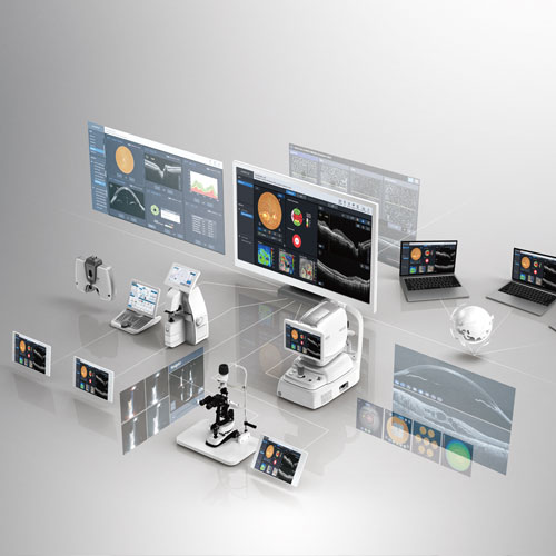
Huvitz Integrated Data Management System
Solution for accurate and intelligent data integration
- Centralizes and manages all measured data in HIIS-1
- Enables analysis based on symptoms and diseases
- Facilitates the analysis of various information
- Offers diverse web compatibility and accessibility
- Support for various data communication
MENU
Explore Details
Centralizes and manages all measured data in HIIS-1
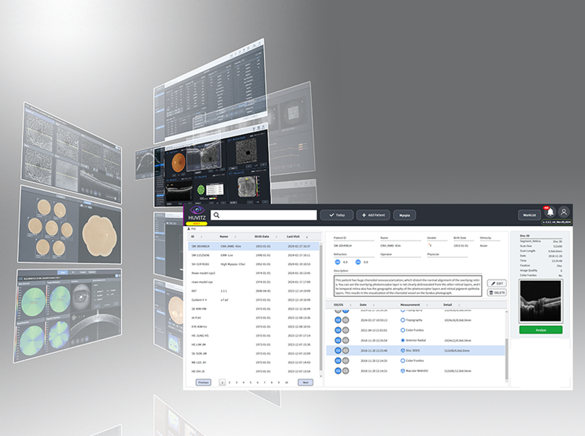
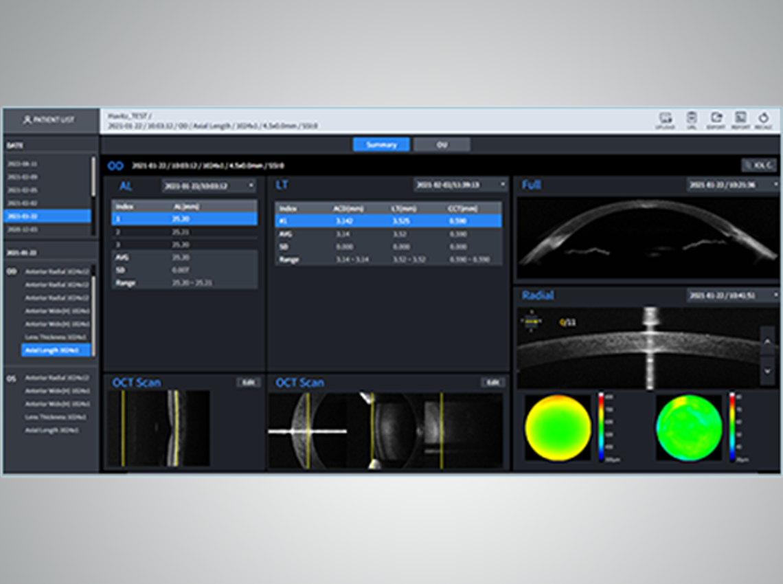
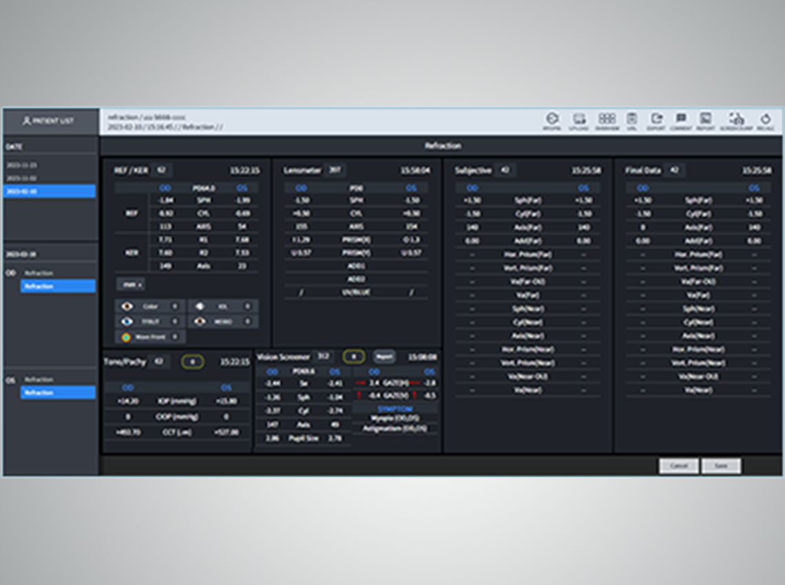
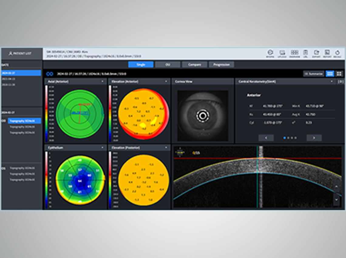
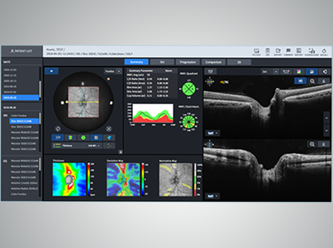
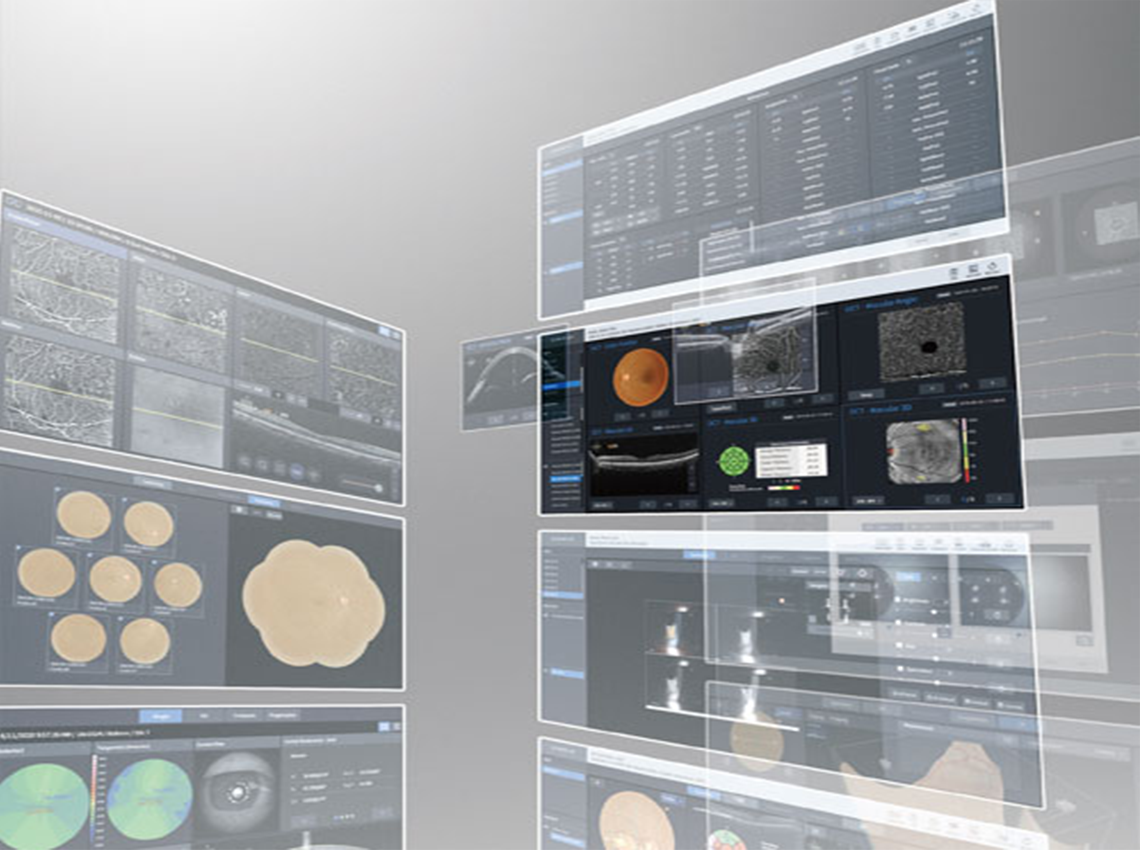
HIIS-1 integrates and manages all data measured by Huvitz products. It organizes data systematically by patient, date, and type of examination, allowing easy access to necessary information. This streamlines the data transmission process and facilitates efficient management of diagnostic results
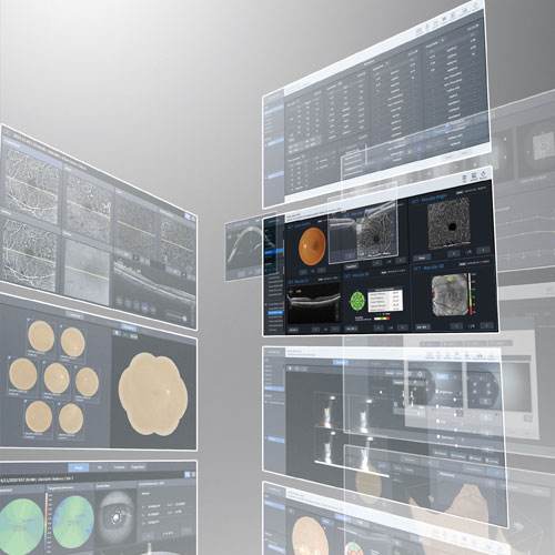
Enables analysis based on symptoms and diseases
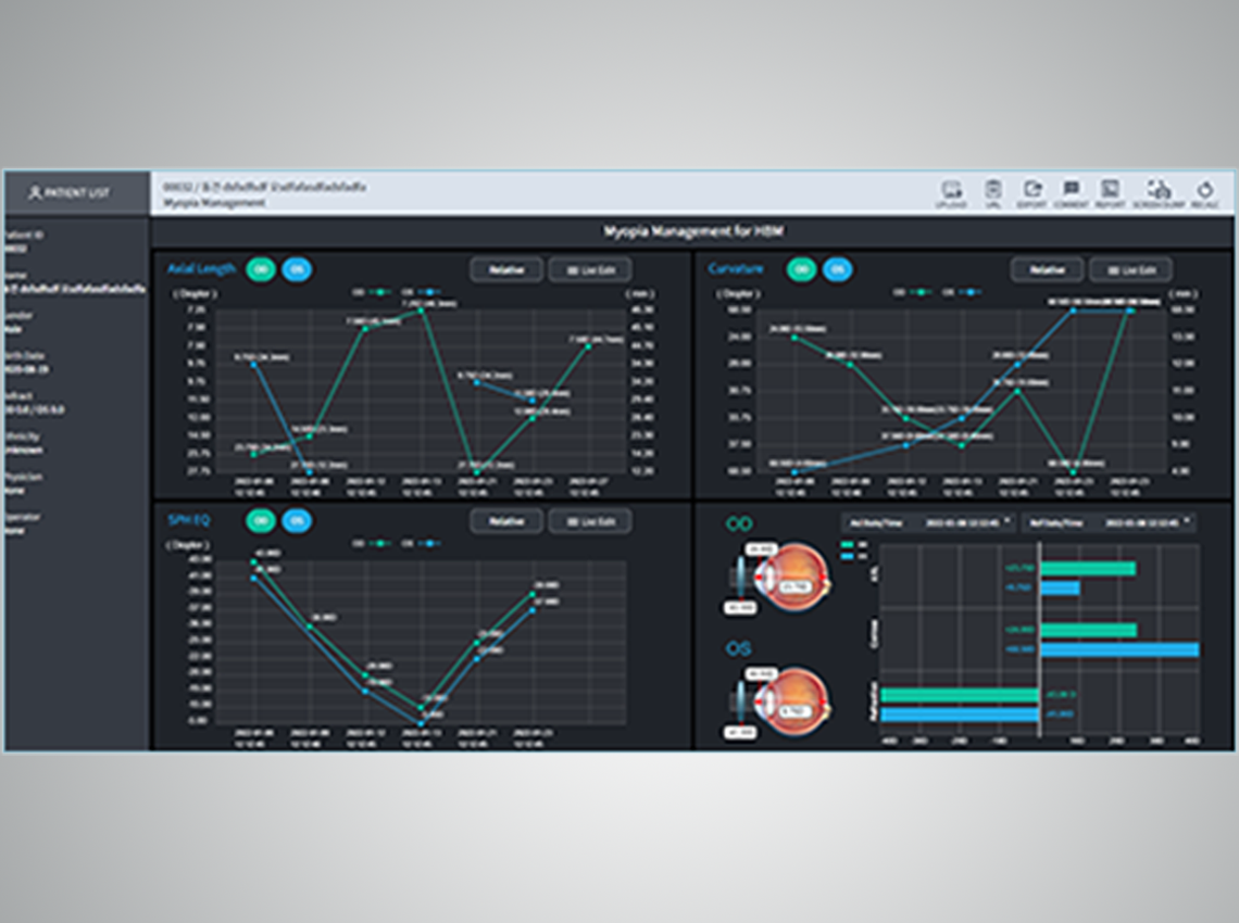
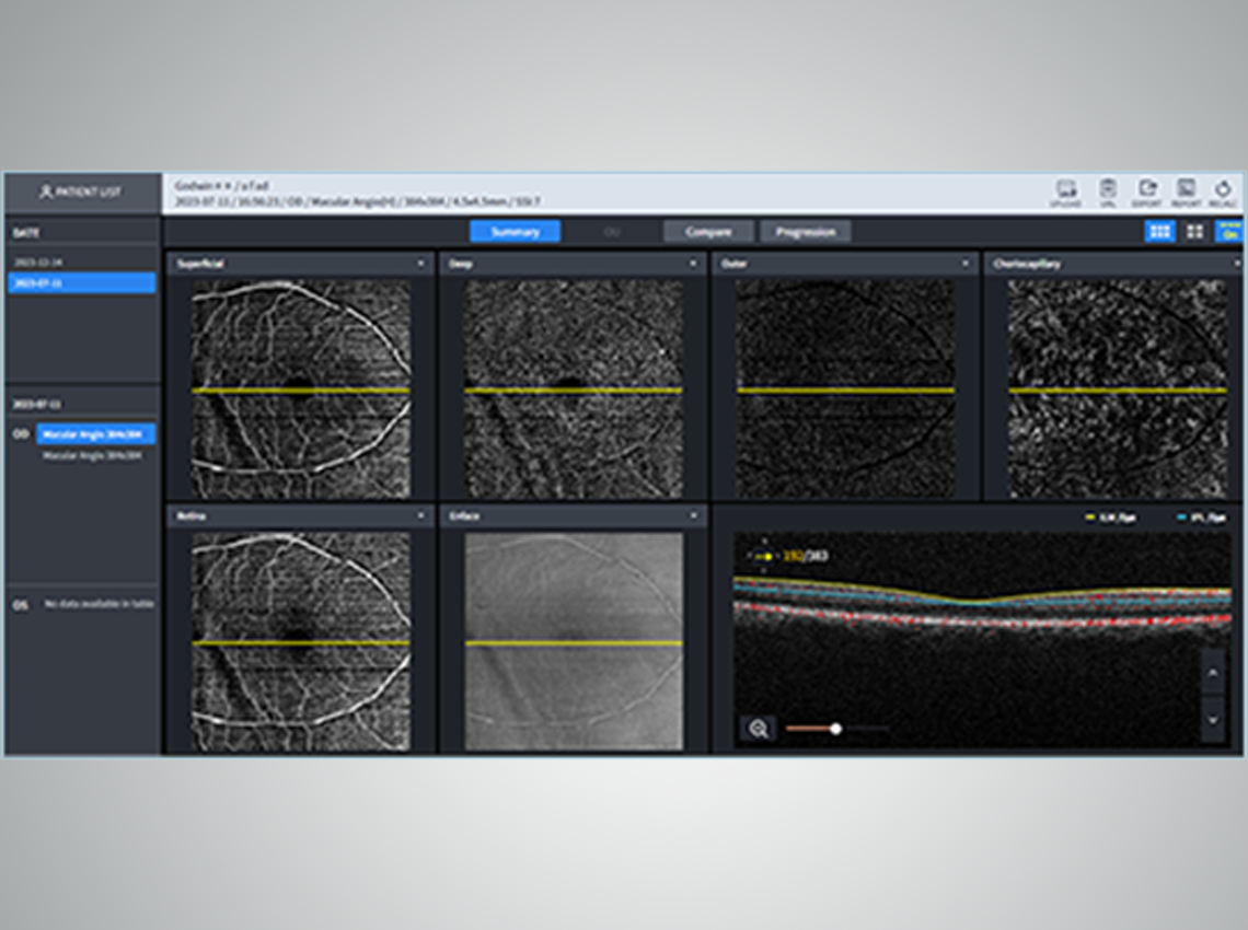
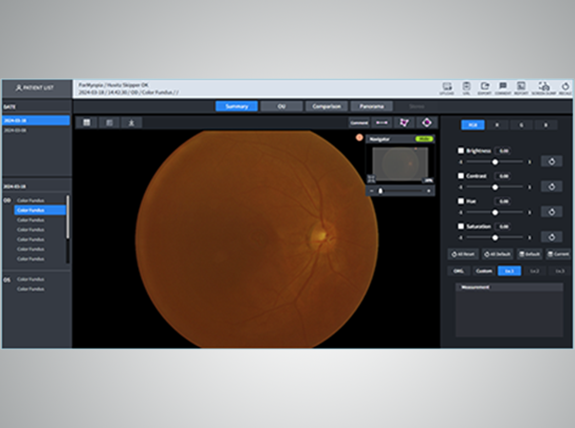
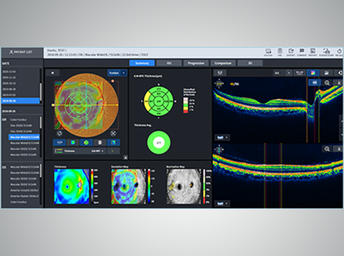
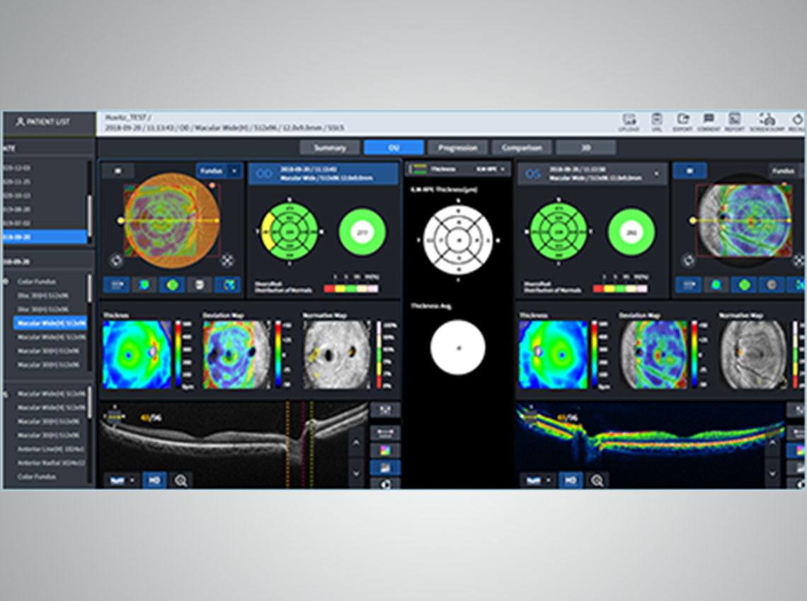
HIIS-1 provides macular, angiography, biometry, topography, and myopia management data depending on the patient’s condition. It offers data based on symptoms and conditions, enabling a more accurate analysis


Facilitates the analysis of various information
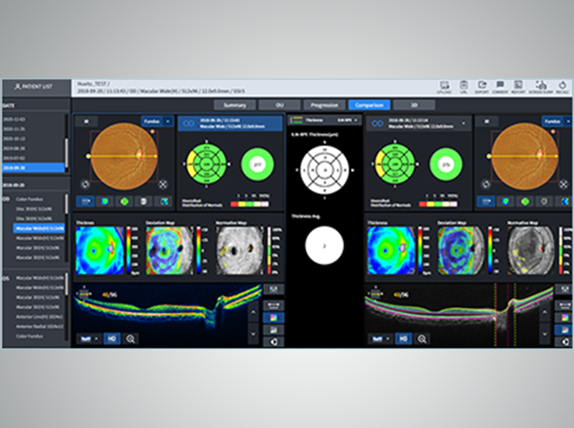
By comparing data between binoculars and analyzing against past data, you can quickly grasp patients’ symptoms, diseases, progression, and past treatment history. This data analysis allows for a clear understanding of the patient’s condition changes and aids in establishing appropriate treatment plans.
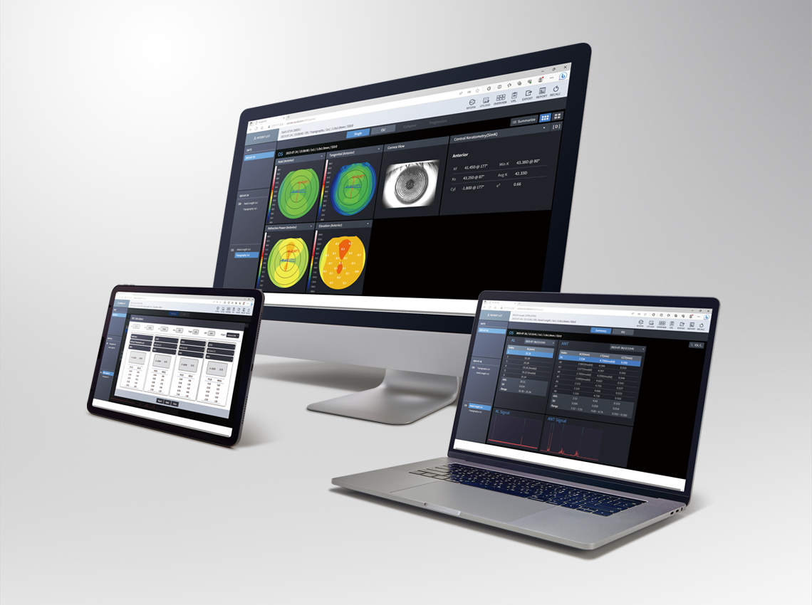
Offers diverse web compatibility and accessibility
You can review patient examination data through a web browser without the need for separate dedicated software installation. Access the data without constraints from various devices such as PCs, mobile phones, and tablets. This allows for convenient access to the patient’s data anytime, anywhere
Support for various data communication
HIIS-1 supports the global standard format DICOM and ensure secure and seamless data integration with electronic medical records (EMR/EHR) systems
Video
Huvitz MEMBERS
If you want more ophthalmology & optometry content
join Huvitz Members
Specifications
1. Type
Web-based software
2. List of supported devices
Refraction
HRK Series, HLM Series HVS, HDR Series
Ophthalmic
HOCT-1/1F, HFC-1, HIS, HNT, HTR-1A, HBM-1, HTG-1
3. External Protocol
Dicom
4. Display
One exam and 6 exams
5. Recommended platform
Server
O/S : Windows 7 or greater (Windows 10 recommended)
CPU : Intel i5 or greater
Memory : 4GB or greater
Ethernet : Fast Ethernet (Gigabit Ethernet recommended)
Client
O/S : Windows 7 or greater (Windows 10 recommended)
CPU : Intel i5 or greater
Memory : 4GB or greater
Ethernet : Fast Ethernet (Gigabit Ethernet recommended)
Browser : Chrome (recommended), IE 11 or greater
6. Feature
Web-Based, Multi users can be accessible
7. Main Function
Communication function:
– Receive patient information, outcome images and data by connecting to ophthalmic medical device productsmanufactured in Huvitz (HOCT-1/1F, HFC-1, HRK-9000A, HLM-9000, HDR-9000,HDR-Mate, HRK-mate, HVS-1, HBM-1, HTG-1, etc.)
Communication function:
– Ability to connect a server with software installed to the web from a client (PC)
Analysis Function:
– Analyzing fundus, OCT and anterior vision images, Angio images
– Refractive power data (Sph, Cyl, Axis) inquiry function measured by the optometry refractive power meter
– Lens refractive power (Sph, Cyl, Axis) inquiry function
– Slit lamp image inquiry function